
Top ▲
GtoPdb is requesting financial support from commercial users. Please see our sustainability page for more information.
Database Links  |
|
| ChEMBL Target | CHEMBL3927 (SARS-CoV) |
| UniProtKB | P0DTC1 (SARS-CoV-2), P0C6U8 (SARS-CoV) |
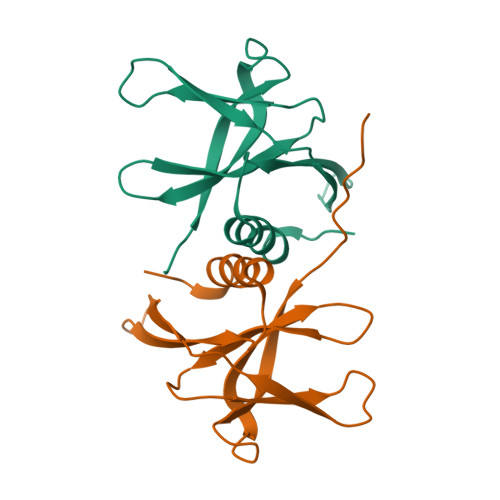
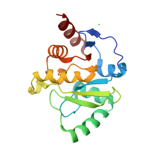
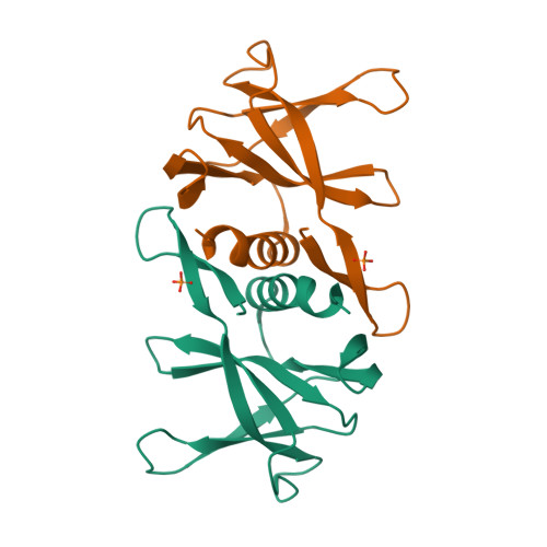
| Selected 3D Structures |
|||||||||||

|
|
||||||||||
|
|
||||||||||
|
|
||||||||||
|
|
||||||||||
| General Comments |
|
The pp1a polyprotein is proteolytically cleaved to form 11 smaller proteins. Most of what is predicted about the functions of these proteins is implied by similarities to orthologues from other coronaviruses [7,15]. Non-structural protein 1: nsp1 binds to host 40S ribosomes and shuts down host translation [9], and effectively blocks RIG-I-dependent innate immune responses that would normally provide anti-viral activity [13]. Non-structural protein 2: Interacts with host prohibitin (PHB) and prohibitin 2 (PHB2) [5] which may compromise mitochondrial respiration and intracellular signalling. Non-structural protein 3 encodes several portien domains, including the papain-like protease (PL-pro) which cleaves motifs at the N-terminus of the replicase polyprotein. PL-pro also removes polyubiquitin chains from cellular substrates, possesses deISGylating activity, and takes part in the assembly of virally-induced cytoplasmic double-membrane vesicles with nsp4. Blocks host IRF3 activation and NF-κB signalling to down-modulate the host anti-viral immune response. Has potential phosphatase activity [11]. An X-ray structure of SARS-CoV PL-pro in complex with an inhibitor compound has been submitted to the RCSB Protein Data Bank (4OW0) [4]. An HCV protease inhibitor, asunaprevir, has been shown to inhibit PL-pro from SARS-CoV, MERS-CoV and SARS-CoV-2 [2]. Nsp3 also contains three tandem macrodomains (Mac), the first of which (Mac1) has ADP-ribosylhydrolase activity. Mac1 proteins from SARS-CoV and SARS-CoV-2 are able to reverse host-mediated mono-ADP-ribosylation by IFN-responsive PARPs, which acts to suppress host IFN production and promote viral pathogenesis. Mac1 has subsequently been explored as a target for anti-coronavirus drug discovery [14]. Non-structural protein 4: Participates in the assembly of virally-induced cytoplasmic double-membrane vesicles with PL-pro [1]. Non-structural protein 5 is more commonly known as 3CL-protease or main protease (Mpro): cleaves motifs in the C-terminus of the replicase polyproteins and is crucial for viral replication. Molecular target for the development of inhibitors with anti-coronavirus activity. Non-structural protein 6: Coronavirus nsp6 proteins limit autophagosome expansion [6]. This mechanism may favour coronavirus infection by damaging autophagosome-mediated delivery of viral components to lysosomes for degradation. Non-structural protein 7: Forms a hexadecameric complex with nsp8 (8 subunits of each) that appears to be involved in viral replication (primase activity) [12]. Non-structural protein 8 Forms a hexadecameric complex with nsp7 (8 subunits of each) that appears to be involved in viral replication (primase activity) [12]. It has been suggested that nsp8 is a second, non-canonical RdRP that generates short primers that are used by nsp12 canonical RdRp for primer-dependent RNA synthesis [8]. Non-structural protein 9 May be a ssRNA-binding protein that contributes to viral replication [10]. Non-structural protein 10 Stimulates nsp14 3'-5' exoribonuclease and nsp16 2'-O-methyltransferase activities, so is essential for viral mRNAs cap methylation [3]. Non-structural protein 11 is only 13 amino acids long and has no confirmed function. |
1. Angelini MM, Akhlaghpour M, Neuman BW, Buchmeier MJ. (2013) Severe acute respiratory syndrome coronavirus nonstructural proteins 3, 4, and 6 induce double-membrane vesicles. mBio, 4 (4). DOI: 10.1128/mBio.00524-13 [PMID:23943763]
2. Anson BJ, Chapman ME, Lendy EK, Pshenychnyi S, D’Aquila RT, Satchell KJF, Mesecar AD. (2020) Broad-spectrum inhibition of coronavirus main and papain-like proteases by HCV drugs. Nature Research, PrePrint, Under Review. DOI: 10.21203/rs.3.rs-26344/v1
3. Bouvet M, Imbert I, Subissi L, Gluais L, Canard B, Decroly E. (2012) RNA 3'-end mismatch excision by the severe acute respiratory syndrome coronavirus nonstructural protein nsp10/nsp14 exoribonuclease complex. Proc Natl Acad Sci USA, 109 (24): 9372-7. [PMID:22635272]
4. Báez-Santos YM, Barraza SJ, Wilson MW, Agius MP, Mielech AM, Davis NM, Baker SC, Larsen SD, Mesecar AD. (2014) X-ray structural and biological evaluation of a series of potent and highly selective inhibitors of human coronavirus papain-like proteases. J Med Chem, 57 (6): 2393-412. [PMID:24568342]
5. Cornillez-Ty CT, Liao L, Yates 3rd JR, Kuhn P, Buchmeier MJ. (2009) Severe acute respiratory syndrome coronavirus nonstructural protein 2 interacts with a host protein complex involved in mitochondrial biogenesis and intracellular signaling. J Virol, 83 (19): 10314-8. [PMID:19640993]
6. Cottam EM, Whelband MC, Wileman T. (2014) Coronavirus NSP6 restricts autophagosome expansion. Autophagy, 10 (8): 1426-41. [PMID:24991833]
7. Fung TS, Liu DX. (2019) Human Coronavirus: Host-Pathogen Interaction. Annu Rev Microbiol, 73: 529-557. [PMID:31226023]
8. Imbert I, Guillemot JC, Bourhis JM, Bussetta C, Coutard B, Egloff MP, Ferron F, Gorbalenya AE, Canard B. (2006) A second, non-canonical RNA-dependent RNA polymerase in SARS coronavirus. EMBO J, 25 (20): 4933-42. [PMID:17024178]
9. Lokugamage KG, Narayanan K, Huang C, Makino S. (2012) Severe acute respiratory syndrome coronavirus protein nsp1 is a novel eukaryotic translation inhibitor that represses multiple steps of translation initiation. J Virol, 86 (24): 13598-608. [PMID:23035226]
10. Miknis ZJ, Donaldson EF, Umland TC, Rimmer RA, Baric RS, Schultz LW. (2009) Severe acute respiratory syndrome coronavirus nsp9 dimerization is essential for efficient viral growth. J Virol, 83 (7): 3007-18. [PMID:19153232]
11. Saikatendu KS, Joseph JS, Subramanian V, Clayton T, Griffith M, Moy K, Velasquez J, Neuman BW, Buchmeier MJ, Stevens RC et al.. (2005) Structural basis of severe acute respiratory syndrome coronavirus ADP-ribose-1''-phosphate dephosphorylation by a conserved domain of nsP3. Structure, 13 (11): 1665-75. [PMID:16271890]
12. te Velthuis AJ, van den Worm SH, Snijder EJ. (2012) The SARS-coronavirus nsp7+nsp8 complex is a unique multimeric RNA polymerase capable of both de novo initiation and primer extension. Nucleic Acids Res, 40 (4): 1737-47. [PMID:22039154]
13. Thoms M, Buschauer R, Ameismeier M, Koepke L, Denk T, Hirschenberger M, Kratzat H, Hayn M, Mackens-Kiani T, Cheng J et al.. (2020) Structural basis for translational shutdown and immune evasion by the Nsp1 protein of SARS-CoV-2. Science, 369 (6508): 1249-1255. [PMID:32680882]
14. Wazir S, Parviainen TAO, Pfannenstiel JJ, Duong MTH, Cluff D, Sowa ST, Galera-Prat A, Ferraris D, Maksimainen MM, Fehr AR et al.. (2024) Discovery of 2-Amide-3-methylester Thiophenes that Target SARS-CoV-2 Mac1 and Repress Coronavirus Replication, Validating Mac1 as an Antiviral Target. J Med Chem, 67 (8): 6519-6536. [PMID:38592023]
15. Yoshimoto FK. (2020) The Proteins of Severe Acute Respiratory Syndrome Coronavirus-2 (SARS CoV-2 or n-COV19), the Cause of COVID-19. The Protein Journal volume, 39: 198–216. DOI: 10.1007/s10930-020-09901-4


